Low-Cost Cardiac Ultrasound Phantom
About Ultrasound Phantoms
Ultrasound phantoms are physical models that replicate human tissue properties for imaging training and device testing. Commercial phantoms are widely used in medical education but may cost hundreds to thousands of dollars, limiting accessibility for early learners and low-resource settings.
Low-cost phantoms have gained traction as an alternative, often using simple materials like gelatin, silicone, or latex to approximate acoustic properties. While many inexpensive designs successfully replicate static anatomy, fewer models capture dynamic physiological motion, which is critical for applications such as cardiac ultrasound training.
This project explores the design of a low-cost cardiac ultrasound phantom capable of simulating basic systolic and diastolic chamber motion using accessible materials and simple fabrication methods.
Design Overview
The phantom was inspired by prior <$10 training models and adapted to better represent cardiac motion. The core concept involved creating a fluid-filled chamber embedded in a tissue-mimicking medium that could be manually actuated to simulate contraction and relaxation.
The design consisted of:
- A latex glove chamber acting as a deformable ventricular cavity
- Surgical tubing connected to a syringe for manual pulsation
- Gelatin as a tissue-mimicking bulk material
- Hot glue sticks to simulate rib shadowing artifacts
This configuration produced a deformable, fluid-filled structure that could be imaged using standard ultrasound systems.
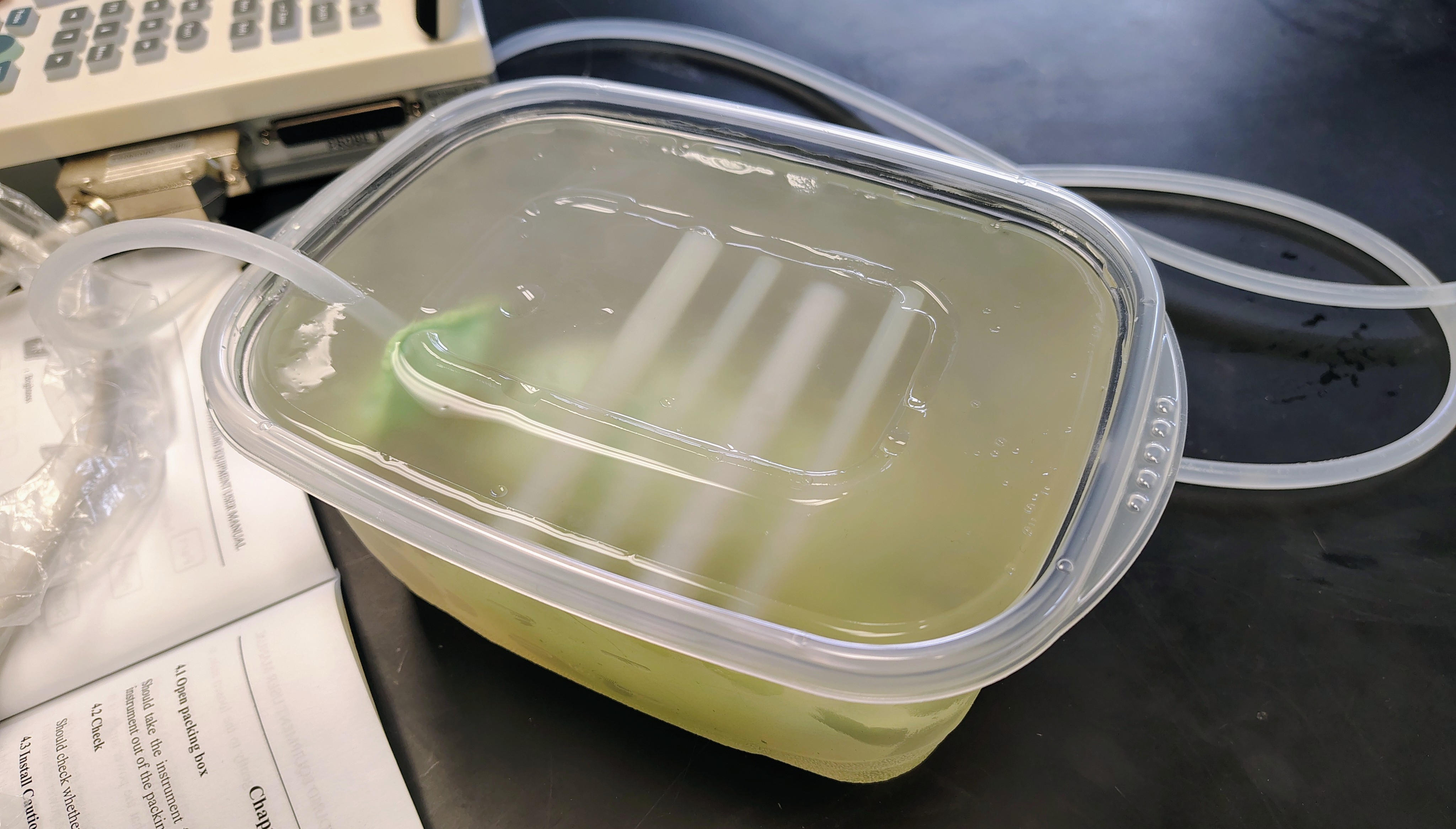
Fabrication
The phantom was constructed by tying off the fingers of a latex glove to form a sealed chamber and filling it with water to mimic a fluid-filled cardiac cavity. The chamber was placed in a plastic container with glue sticks attached to one side, which was then filled with gelatin that had been mixed with calcium chloride for improved structural stability. Tubing connected the chamber to a 20 mL syringe, allowing manual control of internal pressure and cyclic deformation.
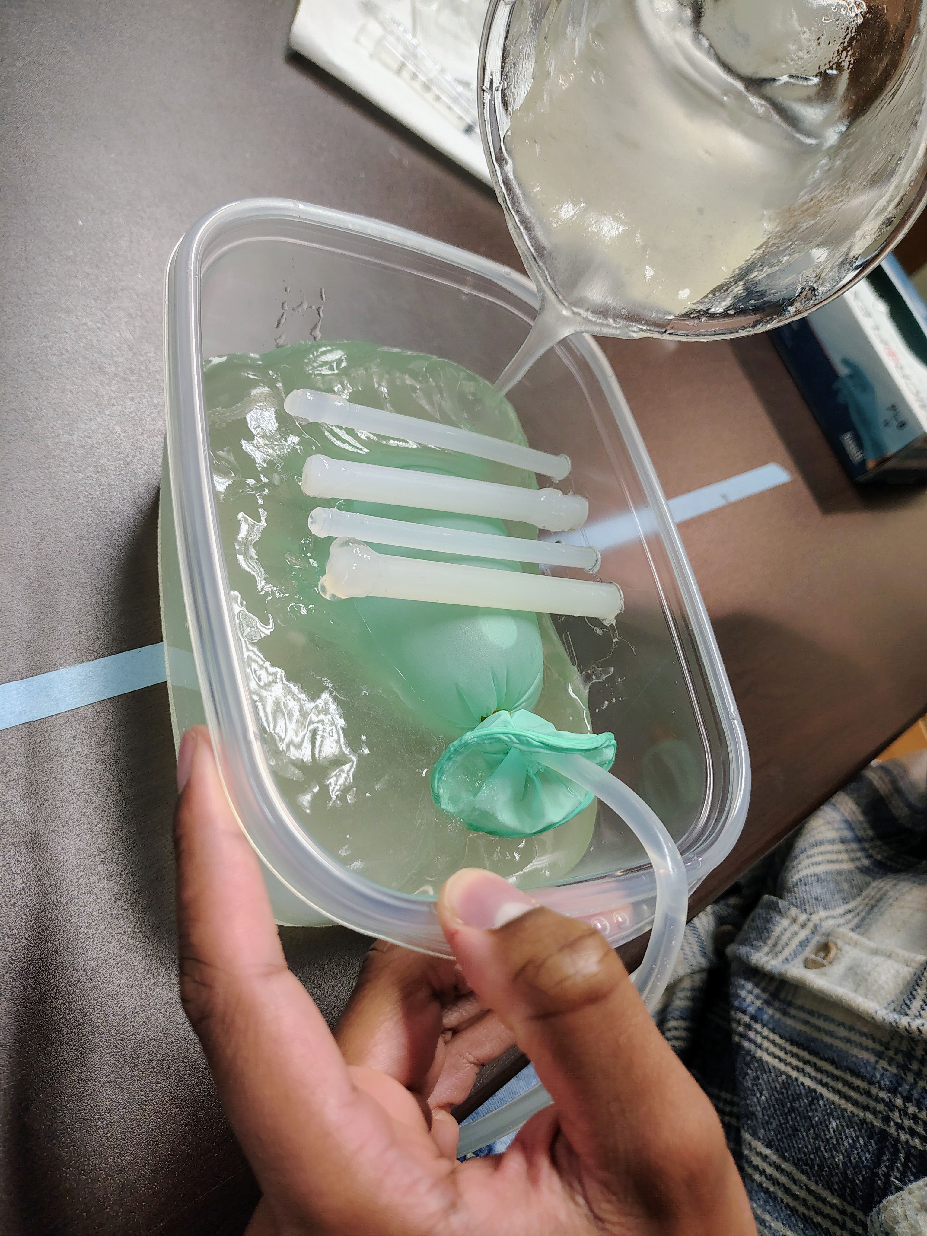
The fabrication process required only inexpensive, widely available materials and was completed in less than 3 hours, making it suitable for resource-limited environments.
Imaging Results
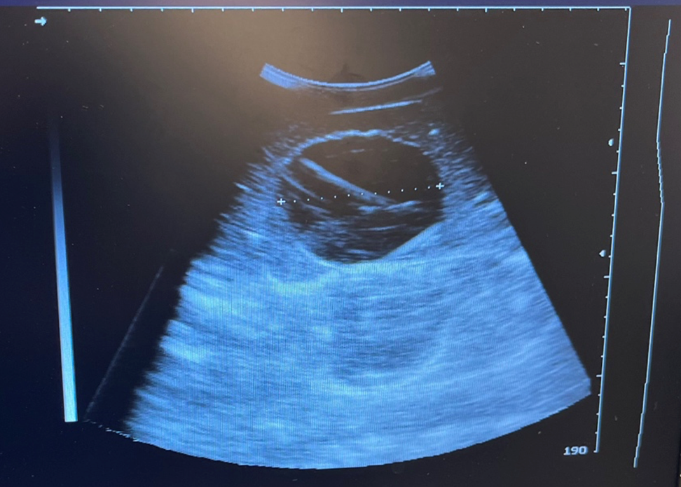
Ultrasound imaging of the phantom successfully reproduced key visual features of cardiac scans, including:
- A clear anechoic chamber resembling a ventricular cavity
- Surrounding echogenic material analogous to soft tissue
- Acoustic shadowing from simulated ribs
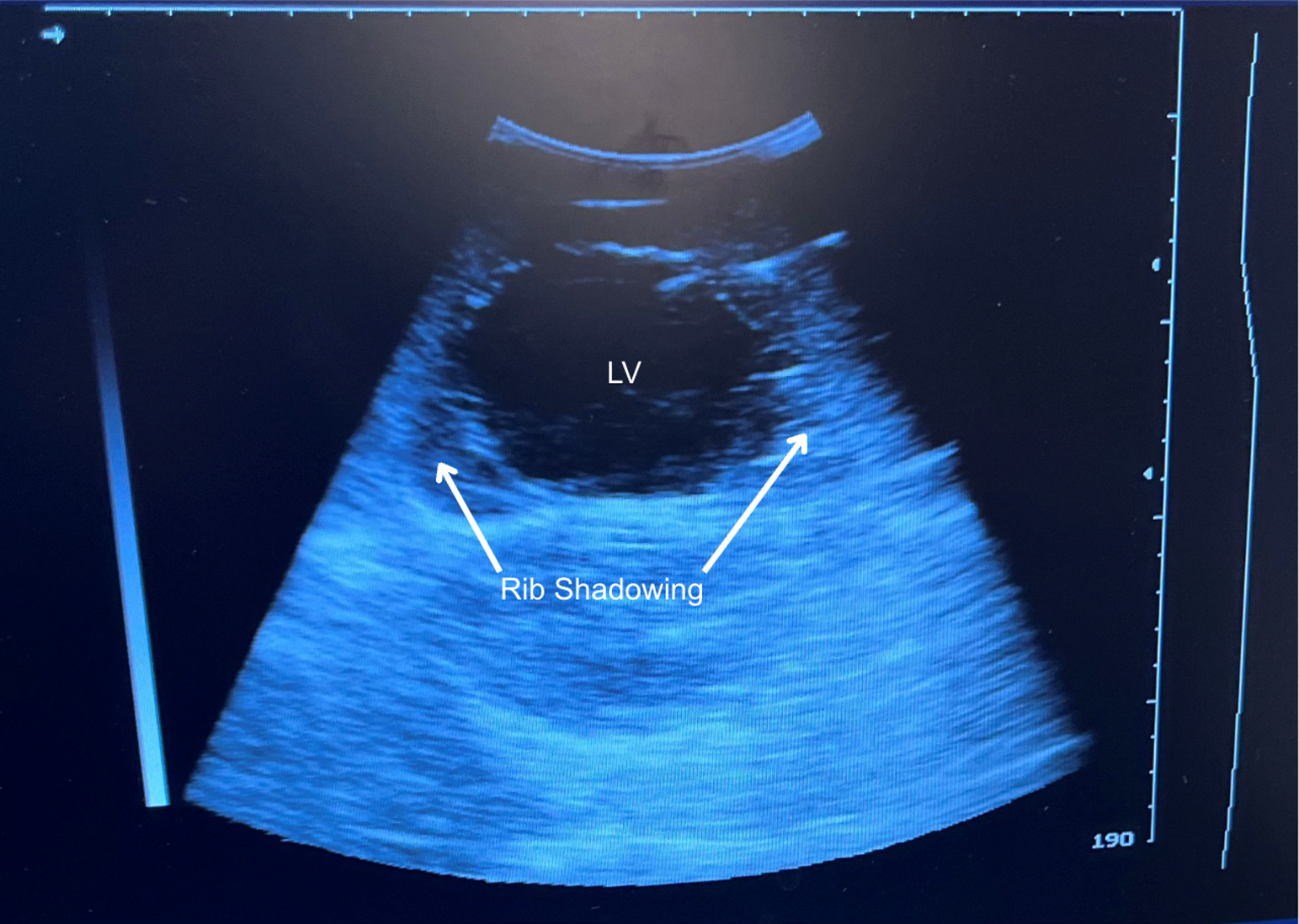
Manual syringe actuation produced visible cyclic deformation of the chamber, enabling visualization of simulated systole and diastole. The chamber diameter decreased from approximately 7.0 cm in the relaxed state to 6.7 cm during compression, demonstrating repeatable dynamic motion detectable under ultrasound.
Discussion
This phantom demonstrates that effective dynamic ultrasound training tools can be produced at extremely low cost. While the model does not replicate full cardiac anatomy or physiological motion, it provides an accessible platform for introducing students to key imaging concepts such as chamber identification and probe positioning.
Future iterations could improve realism in this design through:
- Motorized or pneumatic actuation for automated pulsation
- Multi-chamber designs for anatomical accuracy
- Improved tissue-mimicking materials for acoustic fidelity
This work highlights how accessible materials and rapid prototyping approaches can expand hands-on medical device education and simulation-based learning.